PROFILE OF THERAPEUTICS
Automated Transepithelial Electrical Resistance (TEER) Measurements Allow for Rapid Screening of the Gastrointestinal Toxicity Profile of Therapeutics
Abstract
Methods
Small intestine (SMI) epithelial cells were harvested from post-mortem donors following IRB approval. SMI cells were seeded onto PermaCell™ high pore density (0.4µm, 1.0x10⁸ pores/cm2) PET membrane cell culture 96-well plates (MatTek Life Sciences; Ashland, MA), raised to the air liquid interface (ALI) and cultured in specially formulated culture medium designed to induce differentiation for 2 weeks. An image of the PermaCell™ 96-well system and a representative H&E stained cross-section of the EpiIntestinal™ tissue model are shown in Fig. 1A-D.

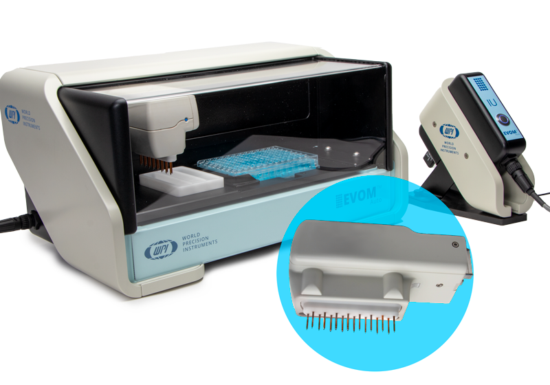
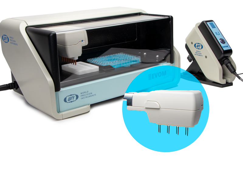
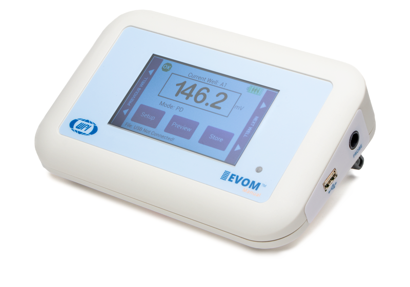
Prior to TEER measurements, the EpiIntestinal™ tissues were transferred to sterile 100 mM KCl (150 µL apical, 400 µL basolateral). TEER was measured both manually using an STX HTS EVOM™ electrode in conjunction with an EVOM™ Manual, and automatically using the EVOM™ Auto automated TEER measurement system (World Precision Instruments, LLC, Sarasota, Florida). A schematic of the Permacell™ 96-well system showing the location of the EVOM™ Auto 96 HTS electrodes is shown in Fig. 1E. A comparison of the EVOM™ Manual with the STX HTS EVOM™ electrode and the EVOM™ Auto systems is shown in Fig. 2A-B. Automated readings were made with a read time of 2 seconds per well. Prior to TEER calculation, tissue sample resistance measurements were background subtracted using the resistance values of blank 100mM KCL buffer in the receiver plate of the 96-transwell assembly. 
Prior to exposure to the test articles, baseline measurements of tissue barrier function (TEER) were taken according to to the methods stated above. Short-term (three-hour) exposure to ethylene glycol tetra-acetic acid (EGTA) was conducted using 1X Hank’s Buffered Salt Solution (HBSS) containing 10mM HEPES as a vehicle applied to the apical surface. Long term (6-day) exposure to dextran sulfate sodium salt (DSS) was conducted by adding DSS to the culture medium in both the apical and basolateral compartments. Reversible tissue damage assessed by TEER is expressed relative to the respective vehicle control tissues.
Tissue samples were fixed for 3 hours in 10% formalin, permeabilized for 30 minutes with 1X Tris-Buffered Saline (TBS) containing 0.1% Triton-X-100 and blocked for 2 hours in 1X TBS containing 10% normal goat serum. Monoclonal recombinant Rabbit anti-Claudin 1 antibody (Abcam cat# ab211737, Cambridge, MA) was diluted 1:1,000 in TBS and incubated at room temp for 2 hours with gentle agitation. Goat anti-rabbit IgG (H+L) secondary antibody, Alexa Fluor™ 555 (ThermoFisher cat# A21429, Waltham, MA) was diluted 1:400 in TBS and incubated at room temp for 1 hour with gentle agitation. Nuclei were counterstained with 4’,6-diamindino-2-phenylindole (DAPI). Images were acquired using an Olympus FV1000 confocal microscope using both a 2X objective and a 40X oil immersion objective. Representative images for IHC staining is shown in Fig. 3D and Fig. 4D for the EGTA and DSS exposures, respectively.

 , where ΔQ/Δt is the concentration of LY present in the basolateral compartment following the time elapsed, Vr is the volume of receiver buffer in the basolateral compartment, A is the surface area of the tissue and C is the initial LY concentration applied to the apical surface of the tissue. D) Immunostaining for barrier protein, Claudin 1 (red), counter stained with DAPI (cyan). Error bars = standard dev.
Tissues were transferred to 250µL of 1X HBSS containing 10mM HEPES and 1.98g/L glucose, and 50µL of 100µM LY dye in the same buffer was applied to the apical surface. The basolateral receiver solution (RS) was replaced every 30 minutes for 2 hours. 100µL from each RS sample was used to determine the LY concentration in the RS at each time point. The RS samples were read using a Spectramax M2 plate reader (Molecular Devices, San Jose, CA) at an excitation bandwidth of 445-455nm, an emission bandwidth of 518-538 nm, and a sensitivity setting of 145.

Conclusions
TEER is a non-destructive analytical technique for functional readout of barrier integrity which can be used to monitor changes to 2D monolayers and 3D tissues models and correlate in vitro and in vivo phenomena.
TEER measurements of the EpiIntestinal™ organotypic tissue model can be used to predict the GIT profile of therapeutics, and more specifically oncology drug-induced diarrhea, a common side effect of oncology drugs which can limit drug utilization.
This study shows the utility of the automated TEER measurements to screen therapeutics on 96-well plates. EVOM™ Auto is an instrument that overcomes the challenges of manual TEER measurements and bolsters the ability to utilize TEER for high-throughput screening.
The use of EGTA and DSS on the EpiIntestinal™ tissue model, can be used to alter barrier function and evaluate a dose dependent response by TEER measurement, Claudin-1 IHC, and Lucifer yellow permeability assay. Compromised barrier function can be detected by TEER measurement and confirmed by LY permeability assay and immunostaining. The withdrawal of the EGTA and DSS treatments recovers the barrier integrity, at least partially, as verified by TEER measurement and LY permeability assay.
We anticipate that the EpiIntestinal™ model together with the automated TEER measurement capabilities of the EVOM™ Auto will be very useful to model normal GI physiology, GI pathology and to screen the potential toxic effects of drug candidates in pharmaceutical development programs.
References
1. Peters, et al., Toxicological Sciences, 2019 Mar 1;168(1):3-17