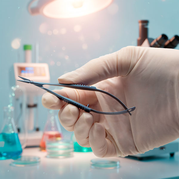
Microscopes
- - July 31, 2025
With various options available for treated or untreated cell culture surfaces, how do you choose the right one for your research? From synthetic polycationic coatings like poly-L-lysine to ECM proteins like fibronectin and vitronectin, each surface treatment offers unique benefits tailored to specific cell specific applications and experimental goals. The choice impacts more than just adhesion. It can influence cell viability, behavior, differentiation, and even affect experimental reproducibility.
In this final post of our series, we’ll summarize the five coatings available on WPI’s our FluoroDish™ glass-bottom culture dishes and help you match the right surface to your application.
- - July 28, 2025
When your cell culture experiments require more than just adhesion, when you need to guide cell behavior, support differentiation, or mimic in vivo tissue structure, fibronectin could be one of the suitable choices.
Fibronectin is a high-molecular-weight glycoprotein found naturally in the extracellular matrix (ECM), where it plays a vital role in cell signaling, migration, and morphogenesis. In vitro, it supports both structural attachment and biochemical communication through integrin-mediated pathways. In WPI’s 35 mm fibronectin coated FluoroDish™ with a 23 mm glass bottom provides a biologically active microenvironment, perfect for small-format, high-qualityimaging experiments where clarity and precision matter.
- - July 25, 2025
Culturing human pluripotent stem cells (hPSCs) requires more than a supportive surface, it demands consistency, control, and clinical readiness. Vitronectin is commonly used for culturing hPSCs since vitronectin supports growth and differentiation of these stem cells.
Vitronectin is an extracellular matrix (ECM) glycoprotein that promotes cell adhesion and survival via integrin binding. It plays a critical role in xeno-free, feeder-free culture systems—especially for labs cultivating embryonic stem cells (ESCs) or induced pluripotent stem cells (iPSCs). WPI’s Vitronectin coated 35 mm FluoroDish™ with a 23 mm glass bottom viewing window, offers a biologically functional, imaging-optimized environment ideal for maintaining stem cells in their most pristine state.
- - June 22, 2025
In most cell culture protocols, improving adhesion plays a critical role, but not every experiment necessarily requires coatings that remain stable long-term or biologically complex substrates. That’s where Poly-L-Lysine (PLL) proves to be a suitable choice
PLL is a synthetic polymer that enhances cell attachment by increasing the surface’s positive charge, helping negatively charged, anchorage-dependent cells adhere more readily to otherwise non-adhesive surfaces like glass or plastic. While it doesn’t mimic the extracellular matrix, PLL remains a trusted choice for labs needing short-term adhesion for cell culture studies of shorter duration, especially during transfection, immunostaining, or fixed-cell imaging. WPI’s 35mm FluoroDish™ with 23 mm glass-bottom culture dishes, provides a consistent, high-clarity platform perfect for observing and documenting cellular events with confidence
- - June 09, 2025
In the world of cell culture, the substrate matters. For many anchorage-dependent cells, simply providing a surface isn’t enough. These cells need biological cues that replicate the natural environment of the body to adhere and grow properly. That’s why surface coating of the substrate plays a vital role in the in vitro cell culture for biomimicry in vivo conditions,
In most tissues inside the body, cells normally stay anchored to extracellular matrix (ECM), a porous network of secreted proteins and proteoglycans. The ECM provides structural support to tissues in vivo and helps in retaining secretory factors, such as growth factors which can regulate cell behavior and function through receptor mediated cell signaling. Collagen is one of the most abundant structural proteins in connective tissues and a major component of the ECM. In vitro, it serves as a powerful coating option to promote cell adhesion, spreading, and function, particularly in sensitive or primary cell types. WPI offers...more
- - May 29, 2025
In any successful cell culture experiment, the story starts at the surface. Whether you're working with primary neurons, stem cells, or epithelial monolayers, anchorage-dependent cells rely on the substrate beneath them to survive, adhere, and thrive. The chemical and biological cues provided by the surface can dramatically influence cell morphology, proliferation, differentiation, and even gene expression.
That’s why surface coatings on cell culture dishes plays a critical role in mammalian cell culture-based research. From naturally derived proteins like collagen, fibronectin, and vitronectin to synthetic compounds such as poly-D-lysine (PDL) and poly-L-lysine (PLL), these treatments transform plain cultureware into biologically active environments. The right coating not only supports attachment, but it can help to maintain normal cellular morphology and guide the cell fate. Anchorage dependent cells, when unable to find appropriate surface to adhere to, can undergo programmed or unprogrammed...more
- - December 13, 2024
Petri cell culture dishes like WPI’s FluoroDishes™ are commonly used in laboratories, but they require precision and care to ensure the accurate results of your work. Let’s look at several common mistakes made with cell culture dishes and how you can avoid them.
- - June 17, 2024
Get the highest quality images and video for your research with FluoroDish Cell Culture dishes. Their optical quality glass bottom is as thin as a coverslip, which ensures the least amount of distortions and excellent heat transfer without any of the autofluoresence issues so common with plastic petri dishes.
Choose the style that suits your application. For live cell imaging, embryo research, and life science researchers working with small sample volumes, the 35mm Fluorodish petri dish with a 10mm well (FD3510) is ideal. Researchers working with expensive chemicals or experimental drugs choose the FD3510. They are also an excellent choice for microinjection applications, because they are designed with the lowest access angle for easier insertion of a micropipette during cellular microinjection. Fluorodishes are also available in 35mm (FD35) or 50mm (FD5040) sizes for cell culturing applications. For better adhesion of neurons, try the 35mm Fluorodish that is coated with poly-D-lysine...more
- - August 15, 2023
WPI’s FluoroDish™ cell culture dishes are optimally designed for a wide variety of cell culture and embryology experiments including high resolution imaging, live cell imaging, electrophysiological recordings of fluorescently tagged cells, and microinjection. FluoroDish™ cell culture dishes are far better than standard petri dishes, because they are designed with optical quality glass bottoms, which offer superior imaging using the latest microscopy technology. WPI’s FluoroDishes™ have been cited in over 550 peer-reviewed publications by cell culture laboratories across the world.
- - July 05, 2023
In any laboratory, having key lab supplies is almost as important as having the major equipment. Choosing a reputable supplier of these necessary supplies is as important as having quality laboratory supplies when you need them. WPI wants to be your partner in early drug discovery, and we stock a wide variety of lab supplies, many of which can ship the same business day. Having a variety of lab supplies ready to ship makes us a dependable research partner. Here are some of the popular supplies that we keep on hand to meet your needs for your upcoming experiment
- - March 03, 2023
In the process of ‘cell cycle’, cells grow and divide into two genetically identical daughter cells. It is regulated by a complex signaling pathway which keeps cell homeostasis by regulating cell division and DNA duplication1. On the other hand, because cancer cells grow and divide indefinitely out of cell cycle control, anti-mitotic drugs are used to suppress abnormal proliferation of cancer cells2. In particular, Nocodazole is known to be a representative anti-mitotic drug for cancer treatment, and it has the characteristics of disturbing microtubule dynamics during cytoplasmic and nuclear division3,4.
- - March 01, 2023
Cytotoxicity refers to the degree of damage to cells. caused by chemical substances or physical factors. Measuring it through cytotoxicity assay is essential for drug development and biological research. Cells undergo complex signaling pathways that causes various cell death processes such as apoptosis, necrosis, and necroptosis. However, most cytotoxicity assays are measured at the endpoint that makes it difficult to study the dynamic response of cells to drugs.
In this application note, we aimed to examine the performance of a cytotoxicity assay using real-time imaging. Cells treated with various concentrations of Nocodazole, the anti-cancer drug, were stained with fluorescent dye during cell death, then monitored with Celloger® Mini Plus. It was observed through time-lapse imaging that apoptosis increased in a Nocodazole dose-dependent manner, and the degree of apoptosis was quantitatively measured and graphed using the Analysis software provided with the Celloger® Mini Plus.
- - February 24, 2023
As white blood cells responsible for immune function are suspension cells that travel along blood vessels, immunology studies often use various suspension cell lines originating from white blood cells. Dealing with suspension cells, unlike adherent cells, slight movement of a plate when locating it on the microscope causes the cells to float. Aside from the problems caused by temperature and CO2 instability, it is in fact not possible to use a traditional microscope to monitor cells in real time. Therefore, in order to stably monitor suspension cells, a live cell imaging device such as Celloger® Mini Plus that operates inside an incubator is essential1. In addition, with Celloger® Mini Plus, the camera inside the system moves to capture the images of cells in multiple positions to keep the cell sample in a steady state instead of having a movable stage with a plate on it. When the suspension cells were monitored both by Celloger® Mini Plus and microscope, imaging with Celloger® Mini Plus...more
- - August 05, 2022
The ATC2000 is a low noise heating system for maintaining animal body temperature during expirimental procedures. Here we show you how to use the adaptive mode on the ATC2000 system.
- Press the Config button to access the configuration menu. Press the Config button again until the adaptive mode Enable displays.
NOTE: If the temperature source is set to heater plate, then the adaptive mode cannot be enabled.
2. Press the Up or Down button to enable or disable adaptive mode.
3. Press the Display button to save the new parameters or press the Config button to toggle through the rest of the parameters.
Once the adaptive mode is enabled, Adaptive appears as the mode on the main display window. If you press the Display button again, the alternate temperature displays along with a timer that counts up from the last sampled time. In this case, the temperature was sampled 5 minutes and 42 seconds ago. If the interval is set to 10 minutes, the temperature will be sampled again in...more
- - April 27, 2022
The ATC2000 is a low noise heating system for maintaining animal body temperature during experimental procedures. The ATC2000 uses a digital PID controller to regulate the application of power to the heating plate to achieve the desired temperature at the monitored sensor. PID stands for Proportional, Integral and Differential. Here’s how PID control works:
The default set point is 37°C, but you can configure it. The controller calculates the error, which is the difference between the present temperature and the desired temperature.
Error = Monitored temperature – Set point
Three individual components are derived by applying different functions to the error.
The default set point is 37°C, but you can configure it. The controller calculates the error, which is the difference between the present temperature and the desired temperature.
Error = Monitored temperature – Set point
Three individual components are derived by applying different functions to the
- The error is multiplied by the proportional...more

Recent Posts