Page 2 - Application Notes
- - April 30, 2013
Stretch receptors are specialized fibers that are in parallel to the fibers in muscle. The receptors stretch as the muscle is lengthened, and they generate action potentials. The frequency of these action potentials is proportional to the length of muscle stretch and the position of the muscle. Through sensory nerve fibers from the stretch receptors, the action potentials and their frequency provide feedback to the central nervous system that will modulate reflex responses and motor control of the muscle.
Setup
For these studies, the whole muscle and the nerve that innervates it are isolated from the organism. A muscle and its nerve, like the Soleus, is isolated and placed in the cuvette of an SI-MT or SI-HTB muscle research system. The muscle is positioned so that the myoneural junction (the place where the nerve innervates the muscle) is on the top side of the muscle. The ends of the muscle are attached to a transducer and a motor through the tissue holders, and the end of the nerve...more
- - April 30, 2013
Abstract
Concentrations of DNA in solution (31µg/mL and 688µg/mL) were measured with a spectrometer and UV/VIS light source in a DIPUV-Mini. Due to the 2mm pathlength, use of a DIPUV-Mini does not require a pre-measurement dilution within this concentration range, thus a potential source of error was eliminated.
Experimental Procedure
Standard solutions of DNA (Sigma D1626) were prepared gravimetrically using 18.2MΩ/cm ultrapurified water as a solvent. Solutions were prepared between 0.0µg/mL and 687.6µg/mL.
Measurements were taken in triplicate using a DIPUV-Mini. The DIPUV-Mini was connected to a UV/VIS light source (WPI #D4H).
Data were collected in 1nm increments across the full range of the instrument (190nm-720nm). The instrument was configured such that reference measurements yielded an 80% total intensity. All measurements utilized 18.2MΩ/cm ultrapurified water as a reference solution.Results
Experimental results are presented in Table 1:
...moreDNA [µg/mL] Absorbance @260nm - - April 30, 2013
Abstract
Concentrations of DNA in solution (31µg/mL and 561µg/mL) were measured with a spectrometer and UV/Vis light source in a cuvette. A 2mm pathlength
cuvette does not require a pre-measurement dilution within this concentration range, thus a potential source of error was eliminated. Although a 2mm cuvette has a total internal volume of 0.7mL, only 350µL is required to obtain an accurate measurement. Experimental Procedure
Standard solutions of DNA (Sigma D1626) were prepared gravimetrically using 18.2MΩ/cm ultrapurified water as a solvent. Solutions were prepared between 0.0µg/mL and 561.1µg/mL.
Measurements were taken in triplicate using a 2mm cuvette (WPI #CUV2102-1) in a standard cuvette holder. An appropriate cuvette spacer (WPI #89342)allowed for consistent placement of the cuvette within the holder. The cuvette holder was attached via two 500µm SMA-terminated fiber optic cables to a UV/Vis light source (WPI #D4H).
Data were collected in 1nm increments across the full...more
- - April 29, 2013
Suction electrodes are used with muscle research systems to record the action potentials from the stretch receptors at the same time that the length and tension of the muscle are recorded.
Materials
- soldering iron with solder
- wire strippers
- wooden-handled dissecting pin
- alcohol burner
- can or tube of contact or plastic cement
- fine flat file
- emery cloth
- electrical tape
- popsicle stick
- two pieces of chlorided silver wire (0.005” dia, 5” long)
(See Chloriding Silver Wire.) - three feet of shielded, two-conductor, insulated cable
- three color-codedconnectors that will mate to the connectors on the input cable for the amplifier
- three feet of flexible plastic tubing (20 gauge Tygon or PE 100)
- 18-gauge needle
- 3-way stop cock
- 3cc syringe
- 1cc tuberculin syringe
- glass micropipette tip
Procedure
- The connectors and electrodes need to be attached to the ends of the shielded, two-conductor cable. Take one end of the cable and carefully strip 5 inches of insulation off the end.
NOTE: Minimize the number...more
- - April 29, 2013
You can use the PZMIV stereo microscope with a stereotaxic frame as shown in the image below. This setup shows a PZMIV-BS. The U-frame Base Plate (502045) is shown, but most stereotaxic frames can be used in this way. Choose a stereo microscope objective that allows you plenty of room to work. For example, the 0.5X objective has 187mm working distance, or the 0.32X objective has 296mm working distance. You could also add a Z-LITE-Z186 illuminator. If necessary, use a 5 to 10 lb.counter weight on the boom stand base to prevent the microscope from tipping.
- - April 28, 2013
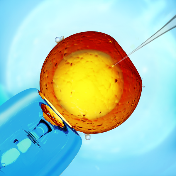
When it comes to setting up microinjection systems, the options appear endless. The pictures below give some broad suggestions on how you might set up your own system. Keep in mind that many parts are interchangeable depending on your needs or preferences.
In general, you will need a stereo microscope on a stand, a light source, one or two micromanipulators with stands, and one or two injection systems. The following images show various setups for microinjection, and all the WPI part numbers are included for easy reference.
Remember, when you set up your own system, choose the parts that fit your needs. For example:
- M10 or the M9 magnetic base could be used.
- PZMIV stereo microscope could be used instead of the PZMIII stereomicroscope.
- M3301 or the KITE micromanipulators can be used, and these micromanipulators can be placed on either side. (Keep in mind, though, if you wanted to use a KITE micromanipulator on the right side of the setup below, you would order a KITE-R (right hand), or...more
- - April 25, 2013
Isolated Stimulation and Stimulus Isolators
The term stimulation refers to the delivery of energy of some kind to a biological tissue in order to elicit an observable response.
Although the energy used in stimulation may be chemical, thermal, mechanical or electrical, this discussion will focus on electrical stimulation. Electrical stimulation of biological tissues involves the delivery of current and voltage to the stimulation site. The two quantities are related by Ohm's law:
V=IR
Where V is the applied voltage, I is the current and R is the electrical resistance of the tissue and or the stimulating electrodes. This simple equation shows that if voltage is constant, current flow will diminish if the tissue/electrode resistance goes up, and will increase if the resistance decreases.
More commonly, the resistance of tissue differs from sample to sample, and the resistance of the electrodes changes with applied current over time in a process called polarization.
Types of Stimulus Devices...more
Recent Posts