Page 15 - Product Information
- - May 16, 2019
Optical measurements are important to researchers. Discover today how WPI has contributed with the recent release of Qualified Fiber.
- - March 12, 2019
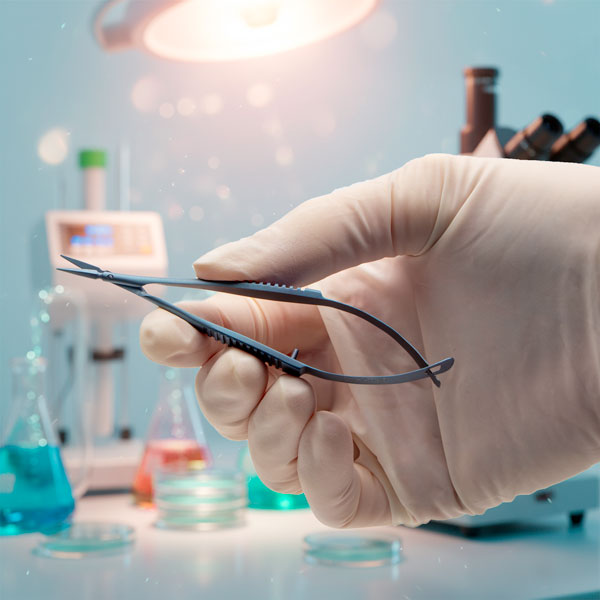
Prior to sterilizing surgical instruments, it is a good idea to make sure you have cleaned them to remove blood, tissue and all other organic material. If soiled materials dries or is baked onto the instruments, it will interfere with microbial inaction and can compromise the sterilization process.
Cleaning is the removal of foreign material (e.g., soil and organic material) from objects and is normally accomplished using water with detergents or enzymatic products.
The most common type of mechanical automatic cleaner is the ultrasonic cleaner. Ultrasonic cleaners are used in conjunction with detergents and enzymatic cleaners. Ultrasonic cleaning removes particulates by cavitation (bubbles) and implosion. Waves of acoustic energy are transmitted in aqueous solutions and disrupt the bonds that hold particulate matter to surfaces.
When selecting detergents or enzymatic cleaners, make sure they are compatible with the metal and other materials of the surgical instrument. The type of...more
- - February 20, 2019
WPI EndOhm chambers are used with WPI's EVOM2 meter for making TEER (transepithelial electrical resistance) measurements. Each EndOhm chamber comes with a "spacer" disk for calibrating the gap between the two chamber electrodes. A consistent gap ensures reliable measurements. Here Subhra shows how to calibrate your chambers.
- - February 20, 2019
WPI EndOhm chambers are used with WPI's EVOM2 meter for making TEER (transepithelial electrical resistance) measurements. After heavy use your background resistance measurement may start to rise. Your electrodes may need to be resurfaced to remove buildup of proteins, sugars and biological materials. Here Subhra demonstrates how to clean and resurface your EndOhm chambers.
- - February 10, 2019
Not all fibers are created equal. When your research is on the line, the choice is clear. Because,WPI optical fibers are qualified for your next project.
...moreThink of it this way–it is optical fiber that finally addresses the lingering issue of fiber performance variability. Qualified fiber ensures repeatability from one fiber cable to the next. Let me explain. Other attributes of a complete fiber optic sensing system have been addressed to ensure repeatability and accuracy from one system to the next. However, given the nature of working with light especially in the UV and with the process of making multimode UV optical fiber, optical fibers have been a limiting factor for product developers. Historically, this critical component of a system WAS an inherent weakness. Not anymore. WPI provides product developers, and researchers with UV basic attenuation data after manufacturing for starters.
The proprietary formulation of WPI fiber resists UV degradation to allow for deep UV down to 185
- - February 07, 2019
Trusted Partner for Reliable Measurements
WPI is dedicated to being your preferred OEM supplier for optical fiber products by offering the following:
- Quality Products and Expert Service – After over 50 years in business, we’ve learned that it takes more to be your partner than just great quality and fair prices. We have implemented measures such as our ISO-9001:2015 certification throughout our home office and production facility to improve our efficiency and ensure quality. We also have invested in six sigma certifications with continuing effort to improve processes within the organization.
- Personal Service – A dedicated OEM account manager and a seasoned optical engineering team with over 20 years of technical design experience make all the difference for our customers.
- Cost Effective Solutions – Quality optical fiber products at the highest value gives you more confidence for less. Our core values are driven by a desire to empower you to fulfill your scientific ideas.
Proprietary qualified...more
- - November 27, 2018
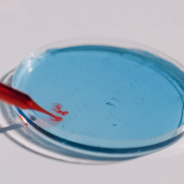
Transepithelial electrical resistance (TEER), also referred as the transepithelial resistance (TER) is used to monitor cellular health. TEER is comprised of measurements of the transcellular pathway (i.e., resistance due to an individual cell) and paracellular pathway (i.e., resistance due to the formation of the cellular junctions). TEER is commonly used to monitor cellular confluence. TEER values can indicate changes in the cellular monolayer permeability, showing the monolayer barrier function of cells such as, endothelial (brain microvessel) and epithelial (alveolar, kidney, and intestinal) cells. High TEER values generally reflect tighter cellular monolayers or cellular junctions (Lewis 1996, Matter and Balda 2003, Denker and Sabath 2011). A few major benefits of WPI TEER measurement systems are described below. The TEER values (electrophysiological analysis) can be combined with other analysis methods to further understand a biological phenomenon. For example, a decrease in the TEER...more
- - October 07, 2018
Measuring CDOM with a Fiber Optic Liquid Waveguide Capillary Cell
One's imagination is the limit with practical uses of WPI's Liquid Wavelength Capillary Cells(LWCC), also referred to in the fiber optic spectroscopy community as a Long Pathlength Flow Cell. This fiber optic sampling accessory for absorbance measurements combines increased optical pathlengths with small sample volumes making them ideal for water analysis such as CDOM.
Two optical fibers couple to this sampling accessory one to send light to the sample cell and the second to send light to a spectrometer. The length of the fiber optic cable can vary allowing investigators to either spot test CDOM in the lab from collected water samples or continuously monitor CDOM of a body of water under investigation with this sampling accessory coupled to a fiber optic spectrometer and source.
What is CDOM?
The optically measurable component of Colored Dissolved Organic Matter. It occurs naturally in water systems and is derived...more
- - August 21, 2018
Clean, Consistent Cut Every Time
When you need to quickly take minimally invasive, small samples, the biopsy punch is an easy choice. The biopsy punch is a hand held, pencil-shaped instrument with a slender, pencil-like body. It is lightweight with a hollow, circular, stainless steel, cutting tip.
In 1887 Edward Lawrence Keyes, the first president of the American Association of Genitourinary Surgeons, was the first doctor who documented the importance of using the biopsy punch for dermatological diagnostics. He observed that the skin tissue samples can be obtained without complications, minimal bleeding and no need for suturing.
Uses for Biopsy Punches
Punches are not solely used in dermatology. Biopsy punches are also designed for therapeutics, cosmetic procedures, and for diagnosing and treating various medical conditions. Punches have found their way into the research world and are frequently used in a variety of applications:
- Electrophysiology–Specimen samples are collected for patch...more
- - July 15, 2018
This 900A video series of 6 videos is designed to help familiarize you with the basic operation of the 900A Micropressure System. The 900A was designed for measuring pressures in kidney tubules, and it has many applications. It has recently been used for measuring ventricular blood pressure in larval fish. We suggest that you become familiar with the information in the instruction manual and the parts and techniques you need to operate the system effectively. Please read the manual and watch these videos prior to setting up and testing your 900A Micropressure System. Instrument Description
- The 900A Micropressure System is designed to measure pressures from –200 to +400 mmHg in small blood vessels, cells and other electrolyte-filled micro-cavities.
- The main components are the 900A include the Control Unit, the Pressure Pod and a Probe that connects to a glass microelectrode.
- The 900A utilizes an ion gradient at the tip of the microelectrode to set a null value of resistance in a...more
- - March 12, 2018
When you are selecting surgical instruments for a procedure, here are a few key points to consider - What procedure are you performing? Published research papers usually indicate which instruments other researchers have used for similar procedures. The correct surgical instrument for a particular procedure makes a difference on the outcome of that technique.
- What is the size of your subject? An instrument that is perfect for a 200–300 g rat (about 22–25 cm long) may not be the best choice for a neo-natal mouse of about 15 g (about 1–2.5 cm long).
- How often will the instrument be used? If you perform more than 100 cuts per day, a pair of titanium scissors or a pair of scissors with tungsten carbide inserts would be worth considering. They stay sharp longer.
In this article we will consider some of these factors and offer a few tips for selecting an appropriate pair of scissors, tweezers and forceps.
Types of Surgical Instruments
Most of our surgical instruments can be used for general surgery...more
- - February 12, 2018
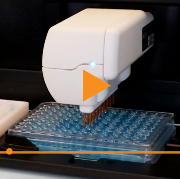
In this video you'll see how to replace the gasket in a Nanoliter2010.
If you have questions, contact us at 866.606.1974 (US Toll Free) or at wpi@wpiinc.com. To request a formal quote, please email us at quotes@wpiinc.com.
- - June 26, 2017
Absorption of light correlates to the energy of a photon that is taken-up by electrons of the substance atom. The electromagnetic energy is transformed into internal energy of the absorbent substance. The absorbance of a substance quantifies how much of the incident light is absorbed by it (instead of being reflected or refracted). Precise measurements of the absorbance at many wavelengths allow the identification of a substance via absorption spectroscopy, where a sample is illuminated from one side, and the intensity of the light that exits from the sample in every direction is measured (see Fig. 1). A few examples of absorption are ultraviolet–visible (UV-Vis) spectroscopy or infrared (IR) spectroscopy.
- - June 26, 2017
The use of fluorescent probes in cell physiology has emerged as indispensable tool in the analysis of cell functioning over recent years. The physics underlying fluorescence is illustrated by the electronic-state diagram (so-called Jablonski diagram, see Fig. 1), showing the three-stage process to create the fluorescent signal (Excitation - Excited/State Lifetime - Fluorescence Emission) in a fluorophore/indicator and simplified described below.
Fig. 1– Jablonski diagram illustrating the processes of fluorescence by absorption of higher photon energy by a fluorophore and subsequent emission of lower photon energy, resulting in fluorescence during the fluorescence-lifetime.
Fluorescence is obtained when an excitation photon (hνEX) from an external source, such as a high-power LED, is absorbed by a fluorophore that elevates its energy (S1’). During the fluorescence-lifetime, the elevated energy (S1’) decays to a lower energy state S1. Then, fluorescence results in the emission of a photon...more
- - November 19, 2015
Are you ready to perform experiments involving a cultured cell monolayer? If your monolayer has not reached confluence, you may have holes or gaps in the monolayer that would prevent collection of valid experimental data. When confluence is reached, the electrical resistance across the monolayer peaks and then plateaus. The EVOM2 meter was designed specifically for this application.
The EVOM2 is the ideal meter for use when you are making manual TEER measurements on small batches of cell cultures and do not require a high throughput system.

Recent Posts