Search "teer-evom"
- - July 16, 2024
Transepithelial/Endothelial Electrical Resistance (TEER) qualitatively measures cellular monolayer health and quantitatively evaluates cellular confluence by measuring the electrical resistance across a cell monolayer. It is commonly used to assess the integrity and permeability of cellular barriers, such as epithelial and endothelial cell layers in cell culture plates. Organ-on-Chip platforms are an emerging market for TEER applications. Let’s look at what TEER is and how it is used.
- - May 16, 2025
In barrier model research, the integrity of your cell monolayers isn’t just important—it’s everything. Whether you're studying epithelial transport, drug permeability, or disease modeling, your ability to trust your data hinges on the health of your cultures. That’s why so many researchers rely on Transendothelial/Transepithelial Electrical Resistance (TEER) as a simple, non-invasive, real-time quantitative evaluation of the integrity of your cellular barrier.
- - May 08, 2025
Transepithelial/Transendothelial Electrical Resistance (TEER) is a widely used quantitative technique to assess the integrity of tight junctions in cell monolayers. WPI’s EVOM™ is the gold standard for TEER measurement and is particularly valuable in studies involving drug transport, toxicology, inflammation, and organ-on-chip systems. TEER measurement provides fast and quantitative data and can be used as an efficient and cost-effective method for drug discovery and development. There are certain biological and technical factors that are known to affect TEER readings. This article reviews the key variables that influence TEER outcomes in cell culture studies and how to ensure consistent TEER measurement analysis can be done by minimizing or eliminating the effects of these factors.
- - May 07, 2024
Cell therapies have revolutionized the field of regenerative medicine, offering promising treatments for a variety of medical conditions. Maintaining the safety, efficacy, and consistency of cell-based therapies is paramount for their successful application in clinical settings. Quality control measures play a vital role in evaluating the quality of these therapies, from development to administration. This article explores the current approaches to quality control in cell therapies, including the utilization of functional measurement techniques such as transepithelial electrical resistance (TEER), to ensure the quality and safety of these promising treatments.
- - August 21, 2025
Cell barriers, such as the intestinal epithelium, blood–brain barrier, or corneal endothelium, are critical for controlling what enters and exits tissues. A compromised barrier can lead to disease, while a strong barrier is essential for maintaining health.
Understanding how our body's protective barriers function is crucial for advancing treatments and developing new therapies. For researchers, accurately measuring barrier integrity is vital in fields ranging from drug development to disease modeling. One of the most important tools scientists use to study these barriers is TEER (Transepithelial Electrical Resistance or Transendothelial Electrical Resistance). This powerful measurement technique provides valuable insights into the integrity and function of cellular barriers that protect our organs and tissues. TEER is a gold-standard, non-invasive technique for quantifying the integrity and permeability of cell monolayers grown in culture.
- - September 05, 2025
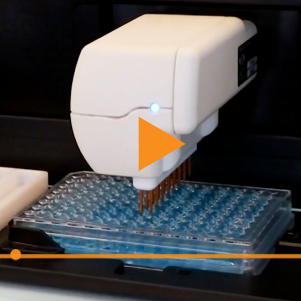
Meet the EVOM™ Auto from World Precision Instruments, the breakthrough that’s revolutionizing drug discovery. When it comes to drug discovery, reliable data on barrier function and cell integrity is essential. Transepithelial Electrical Resistance (TEER) measurements have long been the gold standard for assessing barrier function and cell integrity, critical data for your research, but traditional TEER workflows are often slow, error‑prone, and labor‑intensive.
- - May 03, 2024
Transepithelial electrical resistance (TEER) has emerged as a powerful tool in ophthalmology research and has become a standard method for study of retinal pigment epithelial (RPE) cell biology and the development of these cells as a therapeutic for retinal diseases such as Age-Related Macular Degeneration (AMD). The application of TEER in assessing the barrier function and integrity of RPE cell layers has significantly advanced our understanding of retinal diseases and paved the way for innovative therapeutic interventions including some of the most promising cell therapies to date. This article explores the use of TEER in ophthalmology research, with a focus on studies utilizing RPE cells for both research and clinical applications.
Recent Posts